
You'll want to make sure you get one if you have a family history of congenital heart defects, or if you have diabetes, phenylketonuria or an autoimmune disease. If your doc needs a better listen and view of your baby's heart, he or she may recommend a fetal echocardiogram between 18 and 24 weeks of pregnancy. During your second trimester anatomy scan, your doctor will check the structure of your baby's heart and look for any congenital heart defects. These teeny-tiny blood vessels deliver oxygenated blood to the tissues in your baby's body and then recycle deoxygenated blood back into the circulatory system.īetween 17 and 20 weeks, the heart chambers have developed enough to appear more clearly on an ultrasound. (Up until this point, the cardiac electrical activity occurred spontaneously.)Ĭapillaries also form at an exponential rate during the second trimester. Second trimester developmentīy 17 weeks, the fetal brain has begun to regulate the heartbeat in preparation for life in the outside world. Precursor blood vessels also begin to form in the embryo during the first few weeks. In these early stages, it resembles a tube that twists and divides to eventually form the heart and valves (which open and close to release blood from the heart to the body). At week 5, the preliminary structures that will become your baby's heart begin spontaneously pulsing. First trimester developmentīy week 4, a distinct cluster of cells has formed inside your embryo, which will soon develop into your baby's heart and circulatory (blood) system. How your baby's heart and circulatory system developĮmbryonic heart development starts early on in pregnancy, and your baby's ticker continues to change even after birth as he adjusts from the womb to the outside world.

You could, for example, mistake your own heartbeat for your baby's. Plus, it can be hard to use an at-home Doppler properly without training. That's in part because these devices aren't as sophisticated as the ones that doctors use, so they may not pick up a fetal heartbeat - leading to an unnecessary scare. Trusted Source Food and Drug Administration Avoid Fetal "Keepsake" Images, Heartbeat Monitors See All Sources

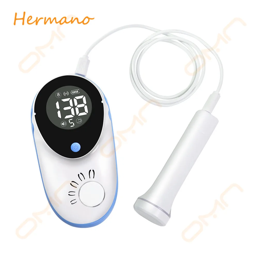
Keep in mind that experts including the Food and Drug Administration (FDA) warn against using at-home fetal Dopplers unless you're under the supervision of a medical professional. Your doctor or midwife will place this handheld ultrasound device on your belly to amplify the pitter-patter of the heart. You'll most likely hear fetal cardiac activity with a Doppler at around the 15-week mark.

When can you hear your baby’s heartbeat with Doppler? Later on, at your 20-week ultrasound (also called the level 2 ultrasound), you'll hear - and see - your baby's heartbeat. Chances are, you'll be able to hear some comforting sounds then. It likely just means that your shy guy is hiding in the corner of your uterus or has his back facing out, making it trickier to pick up on an ultrasound.Īt your next appointment, your practitioner will check to make sure everything is okay. You may see (and/or hear) cardiac activity for the first time from week 6 of pregnancy or later if you have an ultrasound at one of your early prenatal appointments, though the timing of when it can be detected can vary a bit.Ĭan't hear the heartbeat yet? Don't worry. The chambers of your baby’s heart will have developed enough to be seen more clearly on an ultrasound by weeks 17 to 20 of pregnancy. The ultrasound will also confirm your estimated due date, and how many babies you're carrying. If you have a first trimester ultrasound (around or after week 6 of pregnancy), your practitioner or a trained sonographer will check on this embryonic cardiac activity. By week 5 of pregnancy, the cluster of cells that will become your baby's heart has begun to develop and pulse.


 0 kommentar(er)
